Glaucoma Surgery (Trabeculectomy)
Glaucoma affects over 700,000 people in the UK and this number is growing. Thanks to an array of excellent modern treatments, glaucoma is no longer the unavoidable threat to sight that it once was. Careful monitoring of risk factors and early intervention can prevent devastating permanent loss of vision.
Our Videos
Watch our informational videos to find out more about our treatments.

Definition of Glaucoma Surgery (Trabeculectomy)
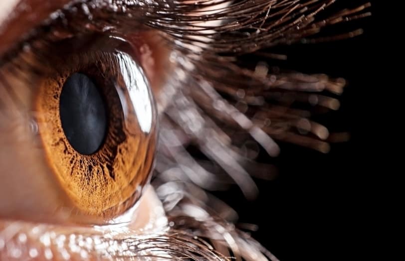
What Is Glaucoma?
Glaucoma is an eye disease that affects the optic nerve. The optic nerve takes the visual information from the eye to the brain, in a similar way that a USB cable downloads images from a camera to a computer. Glaucoma damages this nerve, which results in vision loss.
When the nerve fibres in the optic nerve are damaged, they cannot recover – which also means that any vision lost cannot be recovered. The vision loss usually starts in a patient’s periphery and therefore can go unnoticed for many years till it is too late. For this reason, it is important to undergo regular screening and obtain an early diagnosis to ensure sight is preserved. The good news is that with early diagnosis, regular monitoring and correct treatment, patients have a better chance of maintaining useful vision.
Causes
Causes of Glaucoma
While there is no single cause of glaucoma, there are certain risk factors for the condition, which have a cumulative effect. The most modifiable risk factor is intraocular pressure that is too high for the individual eye to cope with, the numerical value of this varies between person to person.
Other risk factors are:
- Age: The incidence of glaucoma increases as we get older.
- Family history: If you have a family member with this condition, especially a first-degree relative (mother, father, sister, brother or child), your risk of developing glaucoma is far greater.
- Ethnicity: Although certain ethnic backgrounds have a higher risk of developing certain forms of glaucoma, no one ethnicity is immune from its devastating effects. African-Caribbean populations are at higher the risk of primary open angle glaucoma. People of Southeast Asian ethnicity are at increased risk of developing primary angle glaucoma (see ‘Types of glaucoma’).
- Short-sightedness (myopia): Short-sighted (myopic) patients are at higher risk of developing the condition.
- Long-sightedness (hypermetropia): Long-sighted patients are at increased risk of developing angle closure.
- Diabetics: People with diabetes may be at higher risk of developing a secondary glaucoma.
Symptoms
Symptoms and Signs of Glaucoma
Your eyes contain aqueous humour, a clear fluid which helps nourish and protect your eyes whist maintaining its shape. This aqueous humour is flows through the anterior part of your eye producing a constant pressure called intraocular pressure.
In glaucoma, damage to the optic nerve usually occurs if the intraocular pressure rises to an excessive level. This kills the nerve fibres and leads to sight loss. Each individual eye has an optimal intraocular pressure which needs to be maintained.
Some patients can experience glaucoma damage to their optic nerve even though their eye pressure is within the “normal range”: we call this normal tension (or low tension) glaucoma. Conversely, some patients whose intraocular pressure is above the “normal range” won’t go on to experience damage to their optic nerve: this is known as ocular hypertension. Patients with ocular hypertension need to be monitored for signs of glaucoma and may need treatment to prevent glaucoma from developing.
The signs of glaucoma can range from silent and asymptomatic to awareness of a reduction of vision from one or both eyes. It can be painless or associated with discomfort, nausea, vomiting and intermittent visual disturbances
Treatments
Glaucoma Treatment with Sapphire
At Sapphire, our approach to treating glaucoma is centred on patient-specific care, employing a range of innovative therapies tailored to manage this complex condition effectively which is why the best way to tackle glaucoma surgery is by a consultation first.
We understand the critical importance of preserving your vision and offer cutting-edge treatments, from medication management to advanced surgical options, all within a supportive and caring environment. Our dedicated specialists are committed to providing a holistic treatment plan that addresses both the immediate needs and long-term health of your eyes.
Medications (First-line treatment)
- Eye drops are usually the first step
- Prostaglandin analogues (e.g., latanoprost, bimatoprost): increase fluid drainage.
- Beta-blockers (e.g., timolol): reduce fluid production.
- Carbonic anhydrase inhibitors (e.g., dorzolamide, brinzolamide): reduce fluid production.
- Alpha-agonists (e.g., brimonidine): both reduce production and increase drainage.
- Combination drops are available for patients needing multiple mechanisms.
- Oral carbonic anhydrase inhibitors (e.g., acetazolamide) are sometimes used short-term for rapid pressure lowering.
Laser Therapies
- Selective Laser Trabeculoplasty (SLT)
- Increasingly offered as a first-line option and avoids the need for drops (above).
- Targets the trabecular meshwork to improve fluid drainage.
- Can be repeated if pressure rises again.
- Laser Peripheral Iridotomy (LPI)
- Used in angle-closure or narrow-angle glaucoma.
- Creates a small hole in the iris to improve fluid flow.
- Cyclodiode laser therapy
- Reserved for advanced or uncontrolled glaucoma.
- Reduces fluid production by treating the ciliary body
Surgical Procedures
- Trabeculectomy
- Traditional surgery if drops and laser fail.
- Creates a new drainage reservoir (“bleb”) that sits outside the main wall of the eye (sclera) but underneath the skin of the eye (conjunctiva) for fluid to escape.
- Glaucoma Drainage Devices (Tubes/Stents)
- Implanted if trabeculectomy is unsuitable or unsuccessful.
- Includes Ahmed, Baerveldt implants.
- Newer smaller microshunts available which are safer and less invasive than traditional trabeculectomy
- Minimally Invasive Glaucoma Surgery (MIGS)
- Newer techniques (e.g., iStent, Hydrus, Elios, OMNI).
- Lower risk but also less pressure reduction than trabeculectomy.
- Often combined with cataract surgery.
Lifestyle & Monitoring
- Regular monitoring: eye pressure checks, optic nerve imaging, visual field tests.
- Lifestyle adjustments:
- Adherence to drop schedule.
- Avoiding excessive fluid intake in one go.
- Exercise and healthy diet for overall vascular health.
- Smoking cessation and limiting caffeine may help.
- Support: Low-vision services, patient education, and support groups.
Types
Types Of Glaucoma
There are several different kinds of glaucoma: These can be split into primary (no cause for it) or secondary (brought on by another disease process or treatment), open (drainage appears visible but blocked) or closed (drainage is blocked). All managed in different ways, no one treatment fits all.
Primary open angle glaucoma
Open angle glaucoma is the most common form of glaucoma in the UK affecting 1 in 50 adults over the age of 40. Although the drainage angle is open, there is microscopic damage to the trabecular meshwork within the eye, which means the fluid does not properly drain out of the eye. The eye pressure slowly rises, and although it doesn’t cause symptoms in the patient, the optic nerve is slowly being damaged. Damage to the optic nerve results in visual field loss – but you may not be aware of this, since the field loss in one eye may be compensated by the other eye, ‘filling in’ the loss in the visual field. This means that some patients don’t realise they have a problem until extensive damage has occurred. This is why glaucoma is sometimes known as the ‘silent thief of sight’, and is the reason why it’s very important to diagnose and treat glaucoma early.
Primary angle closure glaucoma
In primary angle closure glaucoma, the drainage angle in the eye is ‘occludable’. This means that the aqueous fluid cannot drain through the trabecular meshwork. Since the fluid cannot drain, the pressure in the eye rises. This can happen very quickly, resulting in a very high intraocular pressure and, usually, pain in the eye. The eye can also become red and vision blurred, with halos appearing around bright lights. The pain can be so bad that it causes nausea and vomiting. This condition is known as acute angle closure. It can result in permanent vision loss, in a very short space of time, especially if treatment is delayed. However, if the angle closure attack is treated promptly, the vision recovers.
Sub acute angle closure glaucoma
Sub acute angle closure attacks occur when patients experience mild instances of raised pressure, resulting in blurring of vision, some pain and redness. This then resolves. But if you do experience these symptoms, you should get yourself checked immediately. A more chronic, or slow-developing form of the disease is primary angle closure glaucoma (see above). This is where the intraocular pressure rises to a level that causes damage to the optic nerve but without any noticeable symptoms. The aim of treatment for this condition is to lower the intraocular pressure and open the drainage angle.
.
Normal tension glaucoma
In this condition, the intraocular pressure remains within the normal range (between 10-20mmhg), however, there is still damage to the optic nerve. It is thought that poor blood flow to the optic nerve contributes to its damage. Patients with Raynaud’s Syndrome (a condition that affects blood circulation, causing cold hands and feet) and migraines have a high risk of normal tension glaucoma. This contributes to the theory that blood flow is important in normal tension glaucoma. If blood pressure is too low, it can result in worsening of the disease. For this reason, you may need to have your blood pressure control reviewed. Normal tension glaucoma is treated by lowering the intraocular pressure. This can be done with a number of techniques but usually selective laser trabeculoplasty (SLT) or pressure-lowering drops, in the first instance.
Ocular hypertension
In this condition, the intraocular pressure is raised but there is no damage to the optic nerve and no visual field loss. There is, therefore, no detectable glaucomatous damage. However, since patients with ocular hypertension have a raised intraocular pressure, they are at increased risk of the disease converting to glaucoma, especially if their cornea is thin and the IOP has been under-estimated. These patients may need to be treated to reduce their risk of developing glaucoma or monitored so they can start treatment as early as possible if glaucoma does develop.
Secondary glaucoma
With secondary glaucoma, the raised intraocular pressure does have a specific cause. This might be trauma to an eye, previous surgery, neovascular glaucoma (a condition where blood vessels grow in the drainage angle of the eye because of conditions like diabetic eye disease) or even certain medications like ones used to treat asthma or depression. Here, the glaucoma will need to be treated. The original cause of the glaucoma may need to be treated, too.
Congenital glaucoma
This is a rare condition where developmental abnormalities in the eye cause raised intraocular pressure. Patients may be aware that their vision has always been poor since childhood but may not realise they have an increased risk of further deterioration from glaucoma.
Pricing
Glaucoma Surgery Pricing
Please see below as a guide of prices for glaucoma surgery with Sapphire. For a full list of pricing, please refer to our pricing page here or simply get in touch with our team.
Prices from:
Need help? Simply get in touch with our team.
Prices from:
Trabeculectomy
£4,200
Unilateral SLT
£950
Bilateral SLT
£1,500
Unilateral peripheral iridotomy
£2,000
Bilateral peripheral iridotomy
£2,194
Bleb needling
£2,562
Frequently asked questions
Will I go blind from glaucoma?
Most people with glaucoma don’t go blind, especially if it’s detected early and treated properly. The aim of treatment is to slow or stop further damage. Regular follow-up and using eye drops as prescribed are key to protecting your vision.
Do I have to use my eye drops forever?
In some cases, yes. Glaucoma is a lifelong condition. Stopping treatment usually allows eye pressure to rise again, which can cause further damage. Think of drops like blood pressure tablets – they keep things under control, but don’t cure the condition. There are new alternatives to drops such as lasers and surgery that can take you off your drops permanently.
If my eye pressure is normal, why do I still need treatment?
Glaucoma can cause damage even at normal pressures. What matters is what’s safe for your optic nerve. Your target pressure is usually set lower than average to protect your sight. Treatment is tailored to your specific eye.
Can glaucoma be cured with surgery or laser?
No, there’s no cure yet. Surgery and laser can lower pressure and reduce your need for drops, but they can’t reverse vision already lost. The goal is to protect the vision you still have.
What happens if I forget my drops?
Missing a dose occasionally is unlikely to cause sudden harm, but regularly missing drops increases the risk of sight loss. If you forget, put them in as soon as you remember (unless it’s nearly time for your next dose). It’s best to build the drops into your daily routine.
Can I still drive if I have glaucoma?
Many people with glaucoma can continue driving safely, especially if it’s detected early. The DVLA requires a visual field test to confirm that your vision meets the standards. If your glaucoma affects your driving vision, you’ll be advised accordingly.
Sapphire Eye Care
Ready to learn more or begin your journey?
Simply fill in the form and our team will be in touch.